An illustrated study · Cardiac amyloidosis

The Amyloid Heart.

From misfolded protein to heart failure

identifyamyloid.com

identifyamyloid.com

Drawing of the amyloid cascade: a transthyretin tetramer dissociates into monomers, a monomer unfolds, and misfolded monomers assemble into oligomers and a fibril. A parallel lane shows a plasma-cell clone releasing light chains. Labels mark where each drug class acts: silencers at the liver, stabilizers at the tetramer and depleters, in trials, at the fibril; in AL, an anti-CD38 antibody, a proteasome inhibitor, an alkylating agent and a corticosteroid at the plasma-cell clone.
Drawing of myocardium in long section. Amyloid fills the space between myocytes; a toggle shows Congo red staining in bright field or under crossed polarizers.

Drag to orbit · Scroll or pinch to zoom

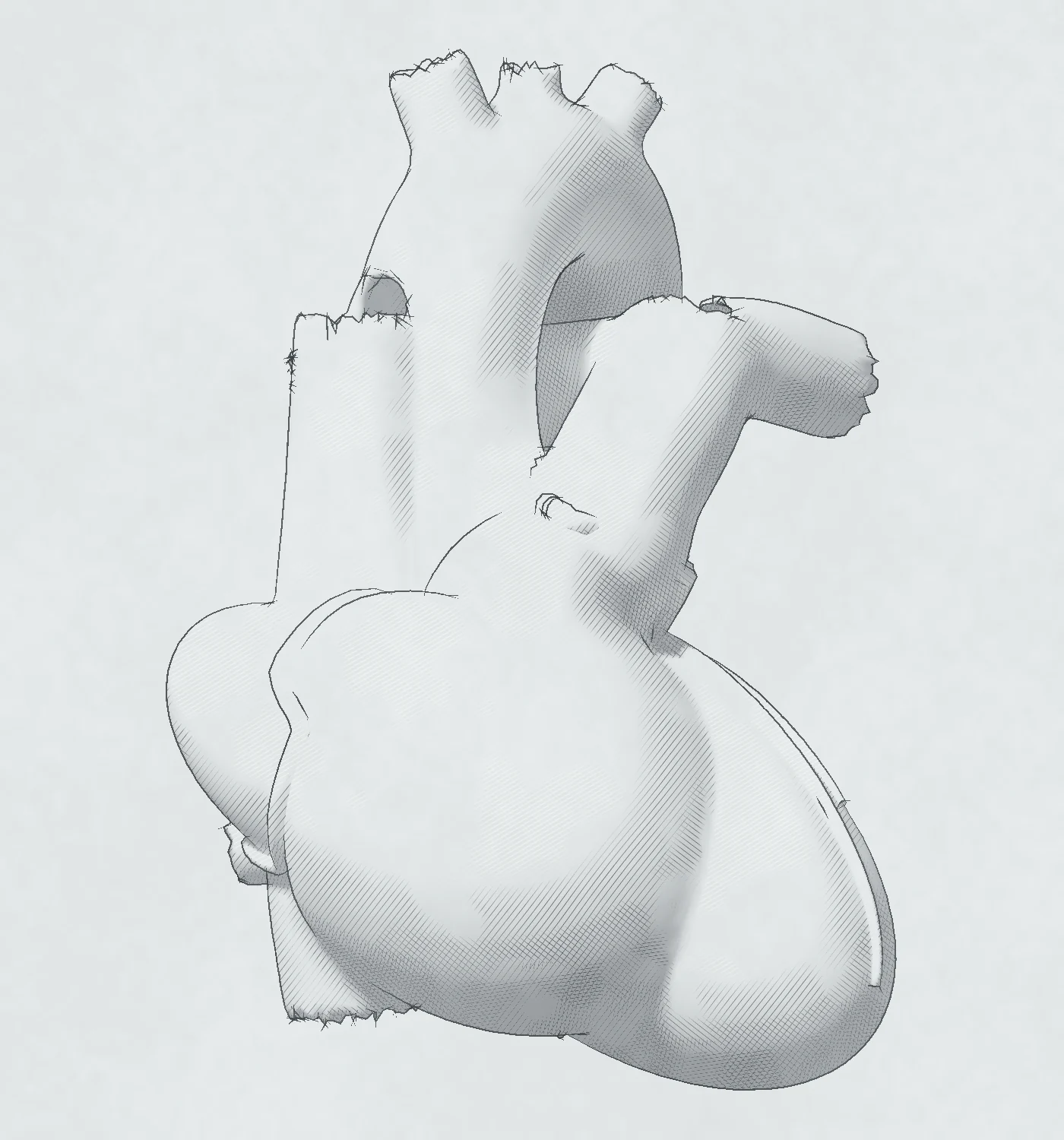
Three-dimensional pencil drawing of the heart as amyloid accumulates. The amyloid burden rises from 0% to 100% and repeats: the ventricular walls thicken, the left ventricular cavity shrinks, both atria enlarge and a pericardial effusion appears.
Left ventricular pressure-volume loop in advanced amyloidosis beside the normal loop: a steep diastolic curve, a high end-diastolic pressure and a small stroke volume.

Imaging plane

Parasternal long axis

Left: the heart model with an ultrasound sector plane cutting through it. Right: the matching echocardiographic view drawn in chalk and graphite, with calipers and numbered red flags.
Mitral inflow pulsed-wave Doppler with a restrictive pattern, septal annulus tissue Doppler with s′, e′ and a′ below 5 cm/s, and a seventeen-segment longitudinal strain bullseye with segmental strain curves showing relative apical sparing, in advanced amyloidosis.
Flowchart of the non-invasive work-up: monoclonal protein screen and bone scintigraphy with SPECT, and the four combinations of results.
Why early diagnosis matters: the hazard of death or heart-failure hospitalization rises 7% for each year from the first heart-failure diagnosis to the ATTR diagnosis; screening finds ATTR cardiomyopathy far more often than routine care, and more often in men than in women; survival falls with the stage at diagnosis. A second view puts the clues on a timeline: carpal tunnel syndrome 5 to 9 years before the diagnosis, first cardiac symptoms more than 4 years before in 42%, the first heart-failure diagnosis a median of 494 days before, and 17 hospital visits in the last 3 years.
How InVision Precision Cardiac Amyloid reads an echo: a segmentation network measures wall thickness on a parasternal long-axis frame, and a video network reads myocardial texture and motion across apical four-chamber frames; the two views give one result per study. A second view compares flags, missed cases and false flags among 1,000 echos for PCA and two simulated sensitivity and specificity pairs.

Misfold

The protein

Two precursor proteins cause more than 98% of cardiac amyloidosis: transthyretin (ATTR) and immunoglobulin light chains (AL).

Type

Transthyretin (TTR) is a 127-residue protein made mainly in the liver. Four chains form a tetramer that carries thyroxine and retinol-binding protein.

The tetramer must come apart before it can misfold. Dissociation into monomers is the rate-limiting step. A partly unfolded monomer then assembles into oligomers and fibrils.

Treatment acts on this chain. Stabilizers bind the two empty thyroxine sites and hold the tetramer together. One stabilizer binds the same sites in a way that mimics the protective T119M variant. Silencers (siRNA or antisense) cut hepatic TTR production. Depleters, antibodies that bind the deposited fibrils, are in a phase 3 trial in ATTR cardiomyopathy.

Three placebo-controlled trials in ATTR cardiomyopathy met their primary outcome:

  • ATTR-ACT: a stabilizer, 441 patients, 30 months. Deaths 29.5% vs 42.9% (HR 0.70); cardiovascular hospitalizations 0.48 vs 0.70 per year.
  • ATTRibute-CM: a stabilizer, 632 patients, 30 months. Win ratio 1.8 on a ranked outcome: death, then cardiovascular hospitalization, NT-proBNP and 6-minute walk.
  • HELIOS-B: a silencer, 655 patients, up to 36 months. Death or recurrent cardiovascular events, HR 0.72; death through 42 months, HR 0.65.

Wild-type ATTR is far more common in men: in the THAOS registry, 94.6% of patients were men. Carpal tunnel syndrome, especially in both wrists, comes 5–9 years before the diagnosis of cardiac amyloidosis. A ruptured distal biceps tendon is another clue.

Variant ATTR is autosomal dominant, with more than 130 known variants. V122I (p.Val142Ile) is carried by about 3.4% of Black Americans and causes a late-onset cardiomyopathy.

In AL amyloidosis, a plasma-cell clone secretes a light chain that misfolds. Lambda outnumbers kappa about four to one, and the heart is involved in about two thirds of patients. The light chains are also directly toxic to the myocardium.

AL treatment targets the plasma-cell clone. The approved first-line combination uses four drug classes: an anti-CD38 monoclonal antibody, a proteasome inhibitor, an alkylating agent and a corticosteroid. The antibody binds CD38 on the plasma cells.

  • ANDROMEDA: the anti-CD38 antibody added to the other three classes; 388 patients with newly diagnosed AL. Hematologic complete response 53.3% vs 18.1%; cardiac response at 6 months 41.5% vs 22.2% of evaluable patients. At a median follow-up of 61 months, death HR 0.62.
  • EMN22: the anti-CD38 antibody alone; a single-arm phase 2 trial in 40 patients with stage IIIB, the group that ANDROMEDA excluded (NT-proBNP above 8,500 ng/L). Survival at 6 months 65%; cardiac response 30%.

The FDA approved the four-class combination for newly diagnosed AL in January 2021. It converted this to traditional approval in November 2025, after the final analysis.

Deposit

The tissue

Fibrils collect between the myocytes. The wall grows thicker, but most of the new mass is protein.

Stain

Amyloid fills the extracellular space. On cardiac MR, the extracellular volume (ECV) of normal myocardium is about 25%. An ECV of 0.40 or more strongly supports cardiac amyloidosis.

In AL, nearly all of the added LV mass is extracellular. In ATTR, the myocytes also enlarge; cell volume rises by about 18%.

The deposits conduct no current, so QRS voltage stays low while wall mass rises. Low limb-lead voltage (0.5 mV or less in every limb lead) was present in 55% of AL and 35% of ATTR patients in one series. A normal ECG voltage does not exclude the disease.

Congo red binds the fibrils. Under crossed polarizers the stain shows the classic apple-green birefringence; in practice several anomalous colors appear.

Remodel

The heart

Thick walls, a small cavity, large atria.

Amyloid burden 0%
View

The LV wall thickens from under 10 mm toward 15–20 mm. The cavity shrinks, so stroke volume falls while the ejection fraction stays near normal.

ATTR often thickens the septum more than the free wall: 79% of ATTR patients had asymmetric septal hypertrophy on cardiac MR, against 14% in AL.

The atria stretch under high filling pressure and are infiltrated themselves. Both enlarge: in 906 patients with ATTR cardiomyopathy, the mean left atrial area was 25–28 cm² and the mean right atrial area 22–27 cm² across three rhythm groups. The interatrial septum, the valve leaflets and the right ventricular free wall thicken too.

About 30% of patients have a pericardial effusion. It is usually small, with a median depth of 7 mm, and rarely limits filling.

Stiffen

The physiology

A stiff ventricle fills fast, then stops.

Amyloid makes the ventricle stiff. The diastolic pressure-volume curve turns steep, so a small rise in volume brings a large rise in pressure. Filling pressure is high although the cavity is small.

Stroke volume is small and fixed, so cardiac output depends on heart rate. In a large ATTR cohort, beta-blockers were stopped in 22% of patients and ACE inhibitors or ARBs in 33%.

The atria fail as pumps even in sinus rhythm, and thrombus can form. An autopsy series found intracardiac thrombus in 33% of hearts, and in 51% with AL.

Early

Why it matters

Amyloid is often found late. Each year of delay adds risk, and a later stage means shorter survival.

Figure

Missed

About 1 in 6 patients undergoing TAVR (16%) had ATTR cardiomyopathy on a bone scan. In heart failure with an ejection fraction of 40% or more and a wall of 12 mm or more, screening found ATTR cardiomyopathy about 5 times more often than it had been diagnosed (6.3% vs 1.3%).

Men carry most of the burden. In the same screening, ATTR cardiomyopathy was present in 10.1% of men and 2.2% of women.

Signs and symptoms

Suspect cardiac amyloidosis in heart failure with a thick LV wall, no clear cause and a non-dilated LV. The clues:

  • Hands and tendons. Carpal tunnel syndrome, especially in both wrists and in men, 5–9 years before the diagnosis. A biceps tendon rupture without trauma.
  • Blood pressure. Hypertension that goes away, orthostatic hypotension, and beta-blockers or ACE inhibitors stopped because the patient does not tolerate them.
  • Rhythm and ECG. Atrial fibrillation; AV block with a thick wall; QRS voltage low for the wall thickness; a pseudo-infarct pattern, seen in up to 70%.
  • Nerves. Neuropathic pain with no clear cause.
  • Diagnoses that hide it. Hypertrophic cardiomyopathy first found after age 60; a troponin rise read as an acute coronary syndrome; low-flow aortic stenosis.
  • AL only. Macroglossia and periorbital purpura. They are specific, but only a minority of AL patients have them, and ATTR patients do not.

Late

42% of wild-type ATTR diagnoses came more than 4 years after the first cardiac symptoms. In the 3 years before diagnosis, patients used hospital services a median of 17 times. In Medicare, the median time from the first heart-failure diagnosis to the ATTR diagnosis was 494 days.

Delay costs

Each year of delay brought a 7% higher risk of death or heart-failure hospitalization: HR 1.07 in Medicare and in the VA, 10,327 patients in all.

Stage at diagnosis

An earlier stage means longer survival. In wild-type ATTR, 4-year survival was 57% at stage I and 18% at stage III; the stage uses troponin T and NT-proBNP. In AL, median survival was 94.1 months at stage I and 5.8 months at stage IV.

Treatment exists for both types.

Image

The echo

Each echo view is a slice through the heart. Here the slice is taken live from the model, so the calipers measure the same walls you see in 3D.

View keys 1–5

Red flags

Measure wall thickness at end-diastole, perpendicular to the LV long axis, at the level of the mitral leaflet tips. Relative wall thickness here is (IVS + PW) ÷ LVIDd.

The subcostal view puts the RV free wall perpendicular to the beam. It also separates a pericardial effusion from the surrounding structures.

Consider

Doppler and strain

Doppler shows how the stiff ventricle fills. Strain shows how each segment shortens: the base stops first, and the apex keeps going.

Figure

Doppler

Relaxation slows first, and E/A falls below 0.8 (grade I). As filling pressure rises, the inflow pattern looks normal again (grade II). It then turns restrictive (grade III), with E/A of 2 or more.

In advanced restrictive cardiomyopathy the deceleration time falls below 150 ms and annular e′ drops to 3–4 cm/s. When septal s′, e′ and a′ are all below 5 cm/s, the tracing shows the “5-5-5” sign.

The atria fail as pumps even in sinus rhythm, so the A and a′ waves shrink.

Strain

Longitudinal strain falls from the base toward the apex. On the bullseye the apex stays red while the base fades: relative apical sparing, often called the “cherry on top”.

Relative apical LS is the mean apical strain divided by the sum of the mean basal and mean mid strain. A value of 1.0 or more separated amyloid from hypertrophic cardiomyopathy and aortic stenosis with 93% sensitivity and 82% specificity in the original study. A later multicenter study found lower accuracy.

The gradient follows total amyloid mass, which is greatest at the base.

An EF ÷ |GLS| ratio above 4.1 flags a normal-looking ejection fraction that hides poor longitudinal function. Normal GLS is near −20%; the lower limit depends on the vendor.

Flag

The AI

InVision Precision Cardiac Amyloid reads the echo the lab already recorded and flags studies suggestive of amyloid.

Figure

Highest specificity. Highest PPV.

InVision Precision Cardiac Amyloid (PCA) has the highest specificity and the highest PPV of any FDA-cleared echo AI for cardiac amyloidosis (FDA 510(k) summaries, cross-study, as of September 2026). In its 510(k) validation, specificity was 99.0% with sensitivity 60.7%. PPV depends on prevalence: 38.4% at 1%, 55.8% at 2% and 76.5% at 5%, where about 3 in 4 flags are amyloid.

Why specificity and PPV matter

Most echos have no amyloid. At 5% prevalence, 950 of 1,000 echos are negative, so each point of specificity is about 10 false flags per 1,000 echos. Each false flag sends a patient to a monoclonal-protein screen and a bone scan.

PPV is the chance that a flag is amyloid. When 3 in 4 flags are amyloid, the work-up goes to the patients who need it. The second figure runs 1,000 echos through PCA and two SIMULATED sensitivity and specificity pairs.

The trade is sensitivity. PCA flags about 6 in 10 amyloid studies, so a study without a flag does not exclude amyloid.

How it reads

PCA reads two standard views with deep learning. On PLAX, a segmentation network finds the walls and measures their thickness. On the A4C clip, a video network reads the texture and motion of the myocardium. Both networks learned from labeled echos, not from hand-set rules, and the two views give one result per study.

Where it fits

  • It reads the echo the lab already records, in adults 65 and over. There is no new test.
  • It is the only FDA-cleared echo-AI amyloid indication with no heart-failure or wall-thickness prerequisite.
  • It is validated in AL and ATTR cardiac amyloidosis.
  • It has FDA Breakthrough Device designation.
  • Its research model is under prospective evaluation at four US health systems (NCT06664866).

After a flag

  1. Review the clues in the history and the ECG, such as carpal tunnel syndrome, QRS voltage low for the wall, or blood pressure that has fallen.
  2. Consider ordering confirmatory testing: a monoclonal-protein screen (serum free light chains, serum and urine immunofixation) and a bone scan with SPECT.
  3. Consider hematology referral if a monoclonal protein is present. In AL, median survival at stage IV is 5.8 months.

For more on InVision Precision Cardiac Amyloid, see invisionmedtech.com.

Diagnose

The work-up

Echo raises the suspicion, by eye or by a flag. Two tests settle most cases.

Bone scan (Perugini grade)
Monoclonal protein

Next two tests

  1. Screen for a monoclonal protein: serum free light chains with the κ/λ ratio, plus serum and urine immunofixation.
  2. Bone scintigraphy with 99mTc-PYP, DPD or HMDP, always with SPECT to confirm that the uptake is in the myocardium and not the blood pool.

Grade 2–3 uptake and no monoclonal protein: ATTR cardiomyopathy can be diagnosed without a biopsy (positive predictive value 100%, 95% CI 98–100). Sequence the TTR gene in every patient, older patients included.

A monoclonal protein is present: refer to hematology and obtain tissue with amyloid typing. A monoclonal protein alone does not mean AL; 5.3% of people over 70 carry one. Conversely, 12% of patients with AL cardiac amyloid show grade 2 or higher uptake.

No uptake and no monoclonal protein: amyloid is unlikely. If suspicion stays high, go to cardiac MR or biopsy; some variants, such as Phe64Leu, take up little tracer.

Perugini grade: 0, no cardiac uptake; 1, mild uptake below bone; 2, moderate uptake with attenuated bone; 3, strong uptake with faint or absent bone. A heart-to-contralateral-lung ratio alone does not make the diagnosis.

Mimics

ConditionWhat separates it
Hypertensive heart diseaseHistory of hypertension. ECG voltage criteria miss most LVH (sensitivity 6.9%), so look for voltage that is low for the wall thickness, and for a patient who is now normotensive.
Hypertrophic cardiomyopathyLVOT obstruction at rest or on exercise in 70%. Asymmetric septal thickening does not separate HCM from ATTR.
Aortic stenosisThe two coexist: ATTR was found in 13–16% of patients referred for TAVR.
Fabry diseaseLow native T1 and basal inferolateral late enhancement on cardiac MR.
HFpEF of other causeScreen the thick-walled patient: 13% of patients aged 60 or more admitted with HFpEF and a wall of 12 mm or more had wild-type ATTR.

Key points

  1. Suspect it in heart failure with a thick LV wall, no clear cause and a non-dilated LV, above all with carpal tunnel syndrome, QRS voltage low for the wall, or blood pressure that has fallen.
  2. Read the echo for it: a wall of 12 mm or more, relative apical sparing, both atria enlarged, a thick interatrial septum and valves, a small effusion.
  3. A PCA flag marks a study suggestive of amyloid. At 5% prevalence about 3 in 4 flags are amyloid, fewer where amyloid is rarer; a study without a flag does not exclude amyloid.
  4. Two tests settle most cases: a monoclonal-protein screen and a bone scan with SPECT. Grade 2–3 uptake with no monoclonal protein diagnoses ATTR without a biopsy; then sequence the TTR gene.
  5. Refer AL now. A monoclonal protein means hematology referral and tissue typing without delay; in AL, median survival at stage IV is 5.8 months.

identify amyloid, with identify struck out and rewritten as InVision amyloid

FDA-cleared AI for echocardiography

For echo labs and health systems

Bring InVision Precision Cardiac Amyloid to your echo lab.

PCA flags echocardiograms suggestive of cardiac amyloidosis in the echos your lab already records, and alerts the interpreting physician for referral to confirmatory testing.

A 30-minute briefing on deployment, reimbursement and the evidence. InVision replies within one business day.

Email [email protected]

Learn more at invisionmedtech.com

Sources & disclosure

Disclosure

InVision Medical Technology Corporation funds and publishes identifyamyloid.com. InVision makes InVision Precision Cardiac Amyloid (PCA).

PCA content
The Flag chapter, the PCA key point in Diagnose and the closing InVision page describe PCA. The other chapters cover the disease, its imaging and its work-up.
Product
PCA (FDA 510(k) K243866) is decision-support software for adults 65+ undergoing echocardiography. It alerts the interpreting physician for referral to confirmatory testing; it does not diagnose. 510(k) validation: sensitivity 60.7%, specificity 99.0%. Rx only.
Claims
Highest specificity and highest PPV: comparison of the FDA 510(k) summaries of FDA-cleared echo-AI devices for cardiac amyloidosis; cross-study, different study populations; PPV compared at equal prevalence. As of September 2026. The two comparison pairs in the 1,000-echo figure are simulated: cases × sensitivity and non-cases × (1 − specificity), rounded.
Audience
US healthcare professionals.
Images
Simulated: the drawings, the heart model, the echo views and the PCA interface. No patient images.
Updated
September 2026
Contact
[email protected] · invisionmedtech.com

© 2026 InVision Medical Technology Corporation

Sources

    The model

    The heart is a parametric teaching model. Signed distance fields describe four chambers, valves, great vessels and pericardium; Blender, driven by script, meshes them and writes one shared topology for four states: normal and amyloid, each at end-diastole and end-systole. The page blends those states per vertex.

    Four-chamber section of the normal model heart, drawn in pencil
    Normal
    Four-chamber section of the amyloid model heart: thick walls stippled red, large atria and a wider pericardial space
    Amyloid

    Four-chamber sections of the same mesh at end-diastole, drawn by Blender (Cycles and Freestyle). Red stipple marks amyloid in the cut wall.

    Measured on the modelNormalAmyloid